IN-2026-015 - Common Cleavers (Galium aparine) - Stem (L.S.) - Epidermis, Vascular Continuity and Pith
Specimen & Context
| Date | 2026-04-02 |
| Species | Galium aparine |
| Common Name | Common Cleavers |
| Material | Fresh stem segment (including internode) |
| Location | Abingdon, Oxfordshire, UK |
| Preparation | Stem, Longitudinal Section (L.S.) |
| Stain | Methylene blue, eosin |
| Series | Scheme of Structural Investigations - Series II — Support and Conduction |
Overview
This investigation examines the longitudinal structure of a herbaceous stem of Galium aparine, with particular attention to the relationship between surface structures, cortical tissues, longitudinal vascular elements, and the central pith.
The aim was to observe the continuity of tissues along the axis of the stem and to identify structural adaptations associated with support and attachment.
The specimen represents a young, flexible herbaceous stem, well suited to examination of axial organisation and surface adaptations.
Method (Summary)
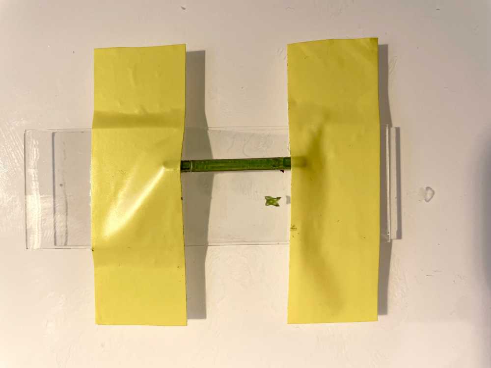
- Electrical tape was used to secure the ends of the specimen to the slide and the slide to the bench, per the above photograph
- This secured the stem during sectioning, to maintain alignment and stability, and left both hands free to take the sections
- A drop of water was added around the exposed portion between the tape
- Longitudinal sections were obtained by controlled, two-handed blade draw along the exposed portion
-
Sections were stained using:
- Methylene blue
- Rinse in water
- Eosin
- Brief final rinse
- Sections mounted in water and examined after staining
Plate Groups
| Plates | Region | Description |
|---|---|---|
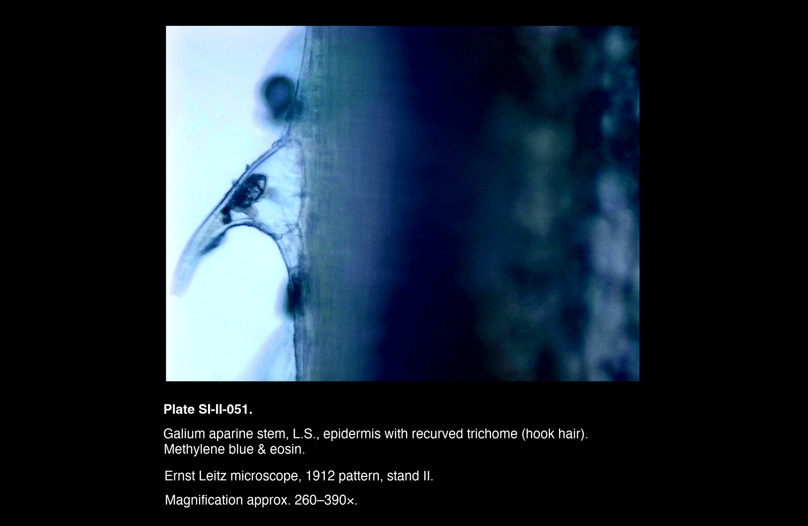
| SI-II-051 | Surface | Epidermis and trichome (hook hair) |
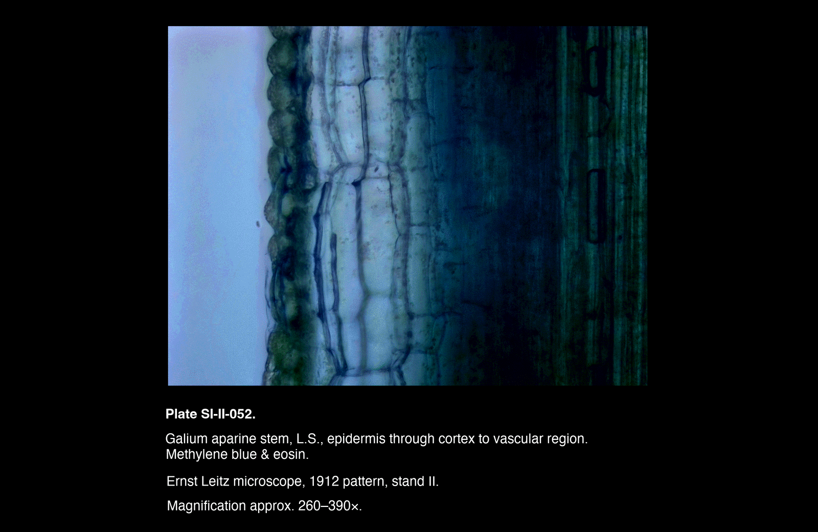
| SI-II-052 | Outer to Inner | Epidermis through cortex to vascular region |
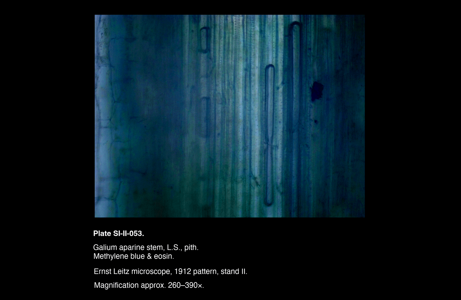
| SI-II-053 | Central | Pith |
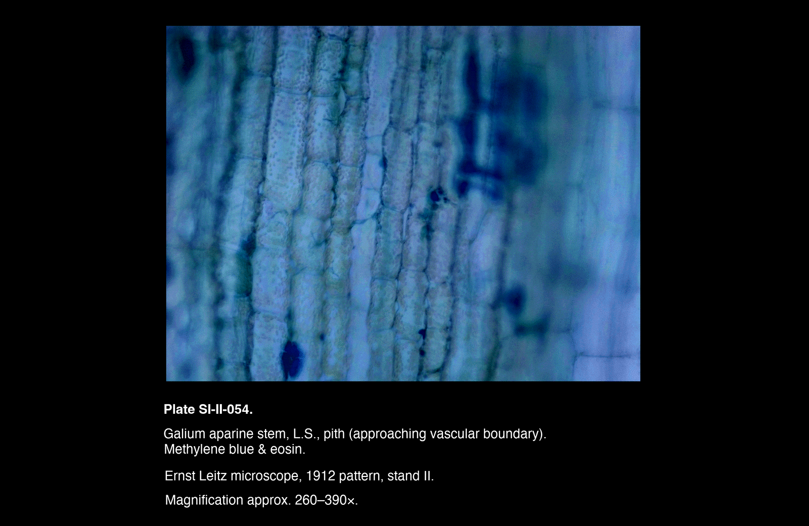
| SI-II-054 | Central | Pith (inner region, approaching vascular zone) |
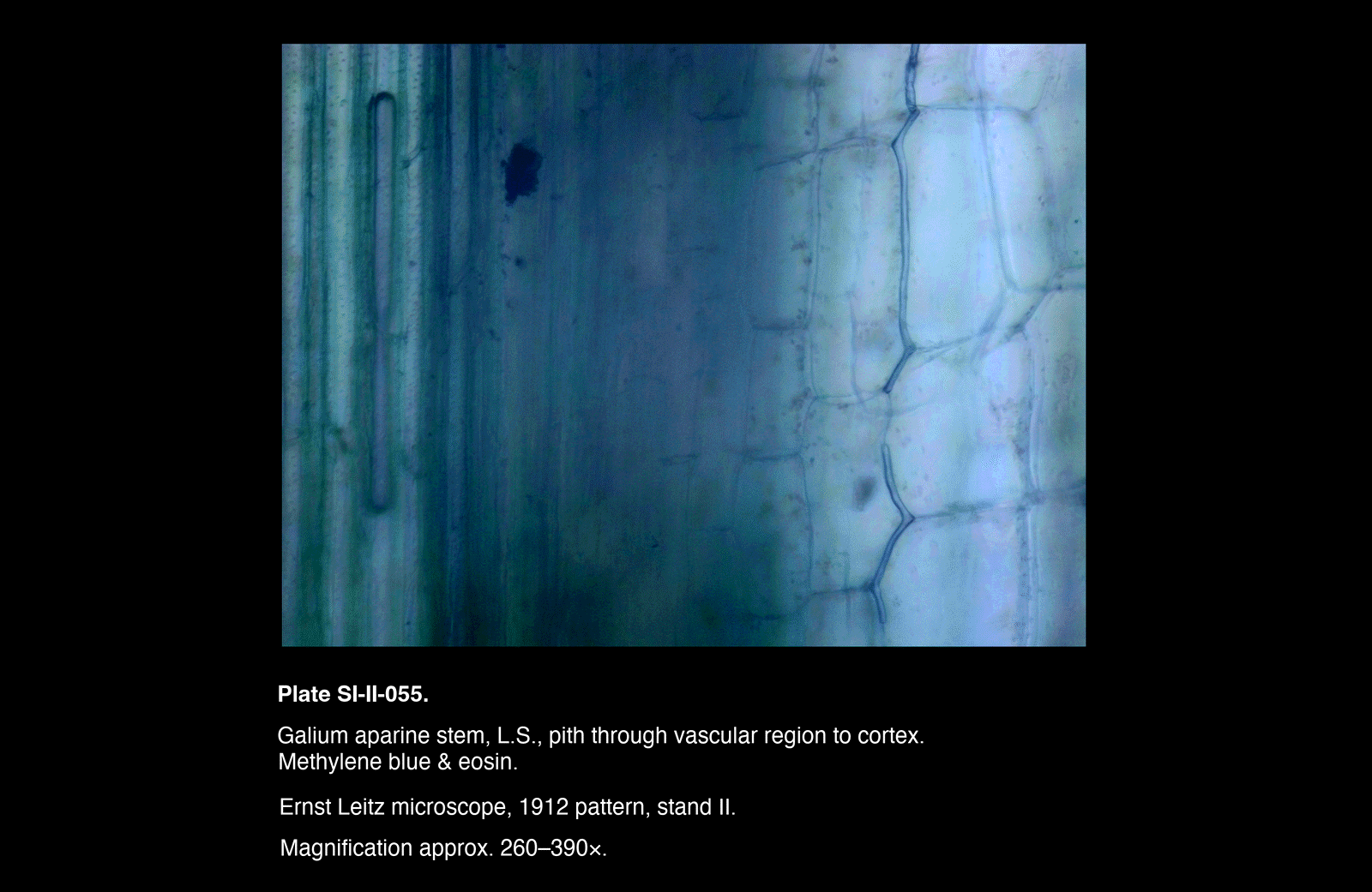
| SI-II-055 | Inner to Outer | Pith through vascular region to cortex |
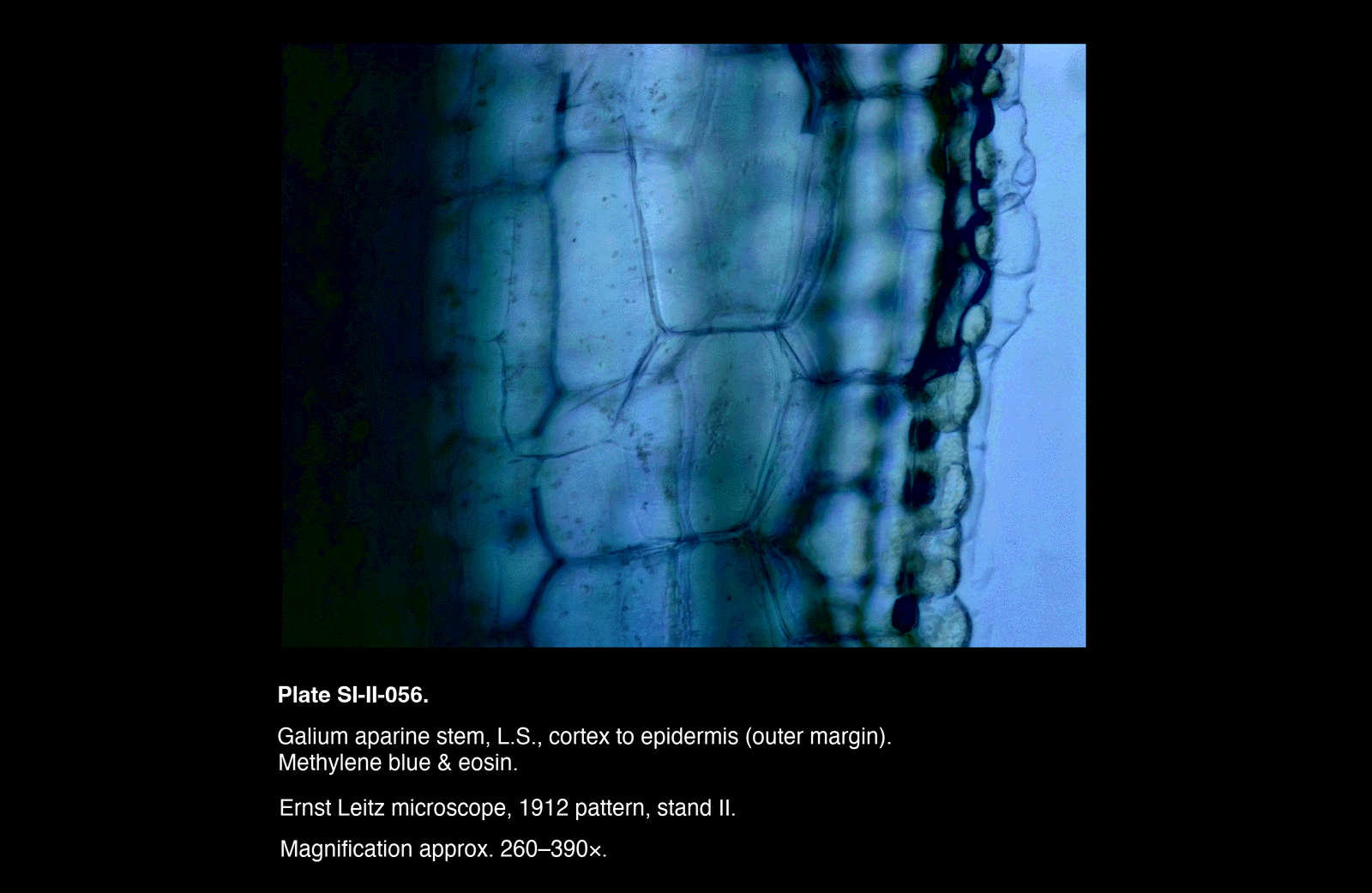
| SI-II-056 | Outer | Cortex to epidermis (outer margin) |
Plates
/Galium%20aparine%20stem,%20L.S.,%20epidermis%20with%20recurved%20trichome%20(hook%20hair))
/Galium%20aparine%20stem,%20L.S.,%20epidermis%20through%20cortex%20to%20vascular%20region)
/Galium%20aparine%20stem,%20L.S.,%20pith)
/Galium%20aparine%20stem,%20L.S.,%20pith%20(approaching%20vascular%20boundary))
/Galium%20aparine%20stem,%20L.S.,%20pith%20through%20vascular%20region%20to%20cortex)
/Galium%20aparine%20stem,%20L.S.,%20cortex%20to%20epidermis%20(outer%20margin))
Plates
Observations
Surface Region (SI-II-051)
- A recurved trichome arises from the surface, showing a curved profile and distinct base
- The trichome is oriented backward relative to the axis of the stem
Outer to Inner Transition (SI-II-052)
Moving inward from the surface:
- Epidermal cells give way to larger cortical cells
- Cortex appears less regularly organised, with broader cell outlines
- A region of denser, longitudinally arranged elements is present deeper within the section
Central Region (SI-II-053, SI-II-054)
- Tissue consists of large, thin-walled parenchyma cells
- Cells are relatively uniform and loosely arranged
- No sharply defined boundary is observed within this region
- In SI-II-054, a gradual increase in structural definition is observed toward one margin
Inner to Outer Transition (SI-II-055)
Moving outward from the central region:
- Open parenchymatous tissue gives way to a denser zone of longitudinal elements
- These elements appear as elongated strands running parallel to the axis
- Beyond this, cortical tissue reappears, with larger, less organised cells
Outer Margin (SI-II-056)
- Cortical cells transition to a more compact outer layer
- Epidermis forms a distinct boundary at the surface
- Cell outlines are more regular and tightly arranged near the exterior
Interpretation
Epidermis and Surface Adaptation
The outermost layer is interpreted as the epidermis, bearing recurved trichomes.
These structures are adapted for:
- Mechanical attachment to surrounding vegetation
- Support of the weak, scrambling stem
The backward orientation of the trichomes is consistent with a clinging function.
Cortical Tissue
The cortex consists of relatively large, thin-walled cells forming a supportive and transitional region between the epidermis and vascular tissues.
Its less ordered appearance reflects its role as a general ground tissue rather than a specialised conducting system.
Vascular Continuity
The denser, longitudinally arranged elements observed in SI-II-052 and SI-II-055 are interpreted as components of the vascular system.
These include:
- Elongated conducting elements (xylem)
- Associated tissues (including phloem)
In longitudinal section, these appear as continuous strands rather than discrete bundles, reflecting their function in axial transport.
Pith
The central region is composed of relatively elongated, thin-walled parenchyma cells consistent with the pith.
This tissue serves:
- Storage functions
- Structural filling of the stem interior
The transition between pith and surrounding tissues is gradual rather than sharply demarcated.
Structural Integration
The sequence of plates demonstrates a continuous organisation across the stem:
- Outer protective and adaptive layer (epidermis with trichomes)
- Intermediate supportive tissue (cortex)
- Longitudinal conducting system (vascular elements)
- Central storage tissue (pith)
This arrangement reflects the integration of support, conduction, and attachment in a herbaceous, climbing stem.
Remarks
- The capture of a recurved trichome in profile (SI-II-051) provides direct evidence of the plant’s clinging adaptation
- The longitudinal disposition of vascular elements is clearly demonstrated, contrasting with their appearance in transverse section
- Intermediate plates (SI-II-053, SI-II-054) are valuable in showing the continuity of pith tissue rather than introducing distinct new structures
- The sectioning method, employing stabilisation of the specimen and controlled blade movement, proved effective in producing consistent longitudinal sections