IN-2026-014 - Hazel (Corylus avellana) - Stem (T.S.) - Periderm and Internal Structure
Specimen & Context
| Date | 2026-04-01 |
| Species | Corylus avellana |
| Common Name | Hazel |
| Material | Fresh twig |
| Location | Abingdon, Oxfordshire, UK |
| Preparation | Stem, Transverse Section (T.S.) |
| Stain | Methylene blue & eosin |
| Series | Scheme of Structural Investigations - Series II — Support and Conduction |
Overview
This investigation examines the transverse structure of a woody stem of Corylus avellana, with particular attention to the organisation of the periderm and the relationship between outer protective tissues, vascular regions, and the pith.
The aim was to identify the principal tissue systems present in a mature stem and to interpret their spatial relationships across the section.
Method (Summary)
- Freehand transverse sections prepared from fresh twig material
-
Sections stained using:
- Methylene blue
- Rinse in water
- Eosin
- Brief final rinse
- Sections mounted in water and examined under consistent optical conditions
Plate Groups
| Plates | Region | Description |
|---|---|---|
| SI-II-048 | Outer | Periderm and surface structures |
| SI-II-049 | Inner | Vascular tissues and pith |
| SI-II-050 | Intermediate | Transition between vascular tissues & pith |
Plates
/Corylus%20avellana%20stem,%20T.S.,%20outer%20region%20showing%20periderm%20and%20surface%20structures.)
/Corylus%20avellana%20stem,%20T.S.,%20inner%20region%20showing%20vascular%20tissues%20and%20pith.)
/Corylus%20avellana%20stem,%20T.S.,%20intermediate%20region%20between%20vascular%20tissues%20and%20pith.)
Plates
Observations
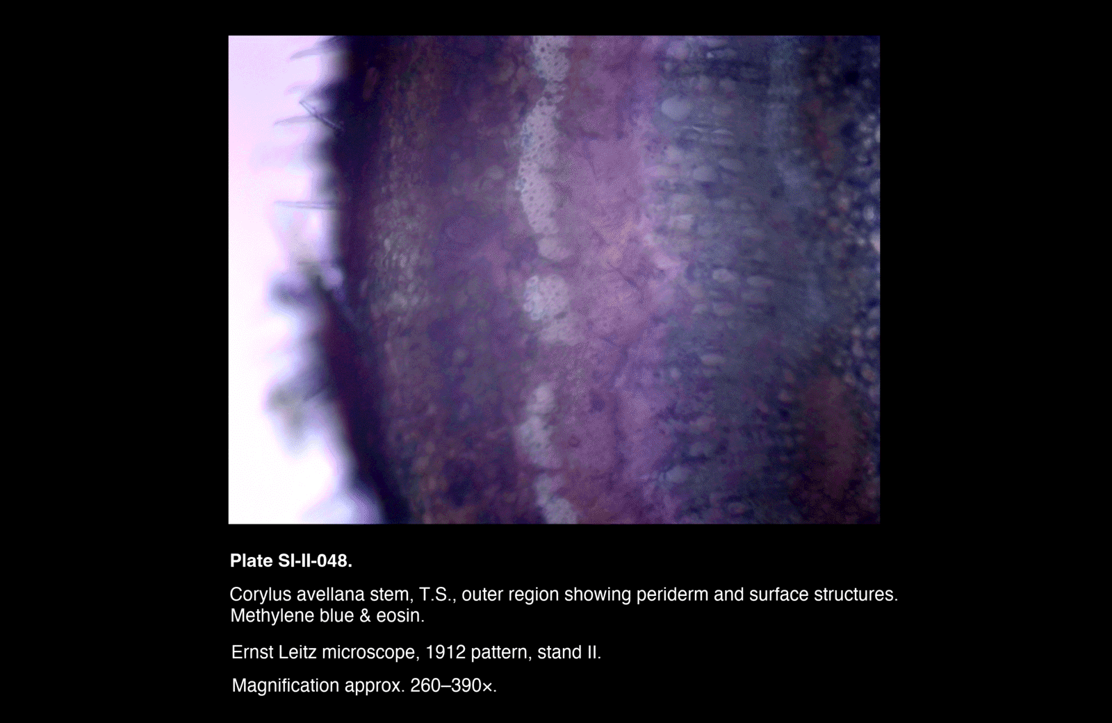
Outer Region (SI-II-048)
Moving from the exterior inward:
- Surface region exhibits projecting hair-like structures consistent with trichomes or their basal insertions
- The outermost boundary appears as a thin, dark band
- Two layers of irregular tissue follow, showing evidence of compression
- A distinct, pale band of relatively uniform cells is present deeper within this region
- Internal to this band, cells become more parenchymatous in appearance
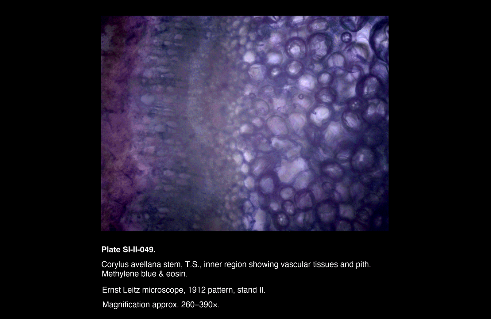
Inner Region (SI-II-049)
Moving from the outer vascular region inward:
- Phloem is present external to a narrow transitional zone
- A thin, poorly differentiated band marks the position of the vascular cambium
- Internal to this, denser tissue consistent with xylem is observed
- Slight inward projections at the inner boundary indicate remnants of primary xylem arcs
- The central region (pith) consists of large, thin-walled parenchyma cells
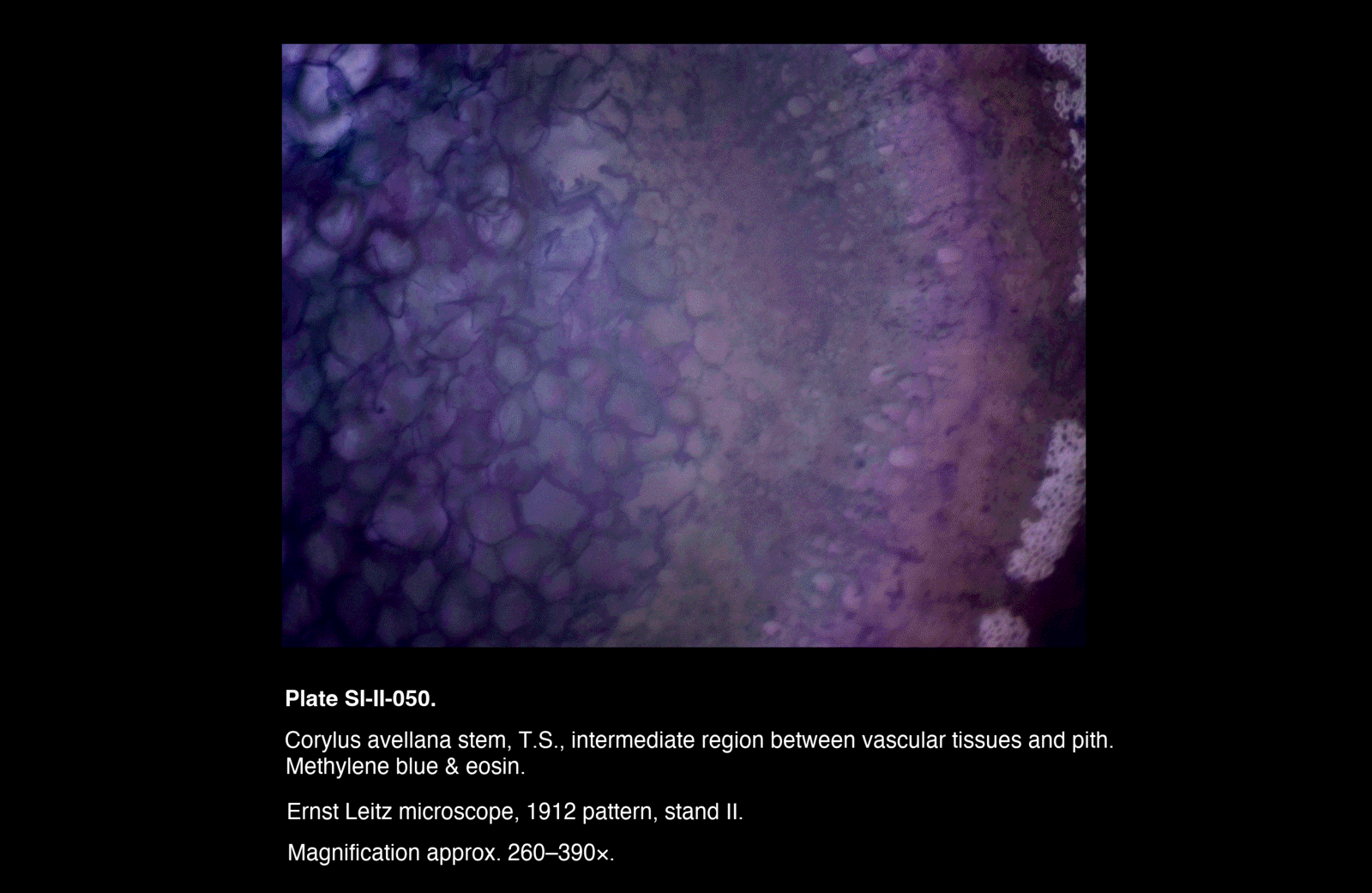
Intermediate Region (SI-II-050)
- Large, thin-walled parenchyma cells characteristic of the pith dominate the field
- Cell size and organisation vary gradually toward denser tissues
- No sharply defined boundary is observed between pith and surrounding vascular tissues
- The region demonstrates a continuous transition rather than discrete layering
Interpretation
Periderm Structure
The outer region (SI-II-048) is interpreted as the periderm, lying beneath the surface layer and outer collapsed cork, comprising:
-
Phellem (cork) Outer, suberised, non-living cells, appearing as darker and often irregular layers
-
Phellogen (cork cambium) A thin meristematic layer, identified as the distinct, pale band of uniform cells
-
Phelloderm Inner living parenchyma, forming a transition toward the vascular tissues
The periderm replaces the epidermis in stems undergoing secondary growth and forms the principal protective layer.
Vascular Organisation
The inner region (SI-II-049) demonstrates the typical arrangement of a woody dicot stem:
- Phloem external to the cambium
- Vascular cambium as a narrow generative layer
- Xylem internal to the cambium
The presence of inward projections suggests remnants of primary xylem, marking the original vascular configuration prior to secondary thickening.
Pith and Internal Continuity
The central pith is composed of large, thin-walled parenchyma cells.
Observations from SI-II-050 indicate that:
- The transition between pith and vascular tissues is gradual
- Tissue organisation reflects developmental continuity rather than sharply defined anatomical boundaries
Structural Integration
Taken together, the plates demonstrate a continuous radial organisation:
- Outer protective tissues (periderm)
- Conductive tissues (phloem and xylem)
- Central storage tissue (pith)
This arrangement reflects the functional integration of protection, conduction, and storage within the stem.
Remarks
- The identification of the phellogen as a distinct pale band proved key to interpreting the periderm
- Variation in outer layers likely reflects successive generations of cork formation and compression
- The intermediate plate (SI-II-050) does not introduce new structures but is valuable in demonstrating continuity between major tissue regions
- The staining protocol provided sufficient contrast to distinguish major tissue systems without obscuring cellular structure