
IN-2026-013 - Red Dead Nettle (Lamium purpureum) - Stem (T.S.) - Staining Comparison
Specimen & Context
| Date | 2026-03-31 |
| Species | Lamium purpureum |
| Common Name | Red Dead-nettle |
| Material | Fresh stem |
| Location | Abingdon, Oxfordshire, UK |
| Preparation | Stem, Transverse Section (T.S.) |
| Stain | Unstained vs. methylene blue & eosin |
| Series | Scheme of Structural Investigations - Series II — Support and Conduction |
Overview
This investigation compares unstained and stained transverse sections of Lamium purpureum stem material using a combined methylene blue and eosin protocol.
The aim was to assess how staining alters visibility of structural features, and to determine whether the method improves interpretability relative to unstained preparations.
The specimen presents clear differentiation between outer tissues, cortex, and vascular regions, making it suitable for evaluating contrast enhancement through staining.
Method (Summary)
- Freehand transverse sections prepared from fresh stem
- Sections initially observed mounted in water (unstained series)
-
Subsequent sections stained as follows:
- Methylene blue
- Rinse in water
- Eosin
- Brief final rinse
- Observations made under consistent optical conditions
Plate Groups
| Plates | Condition | Description |
|---|---|---|
| SI-II-039 to 043 | Unstained | Overview, outer, detail |
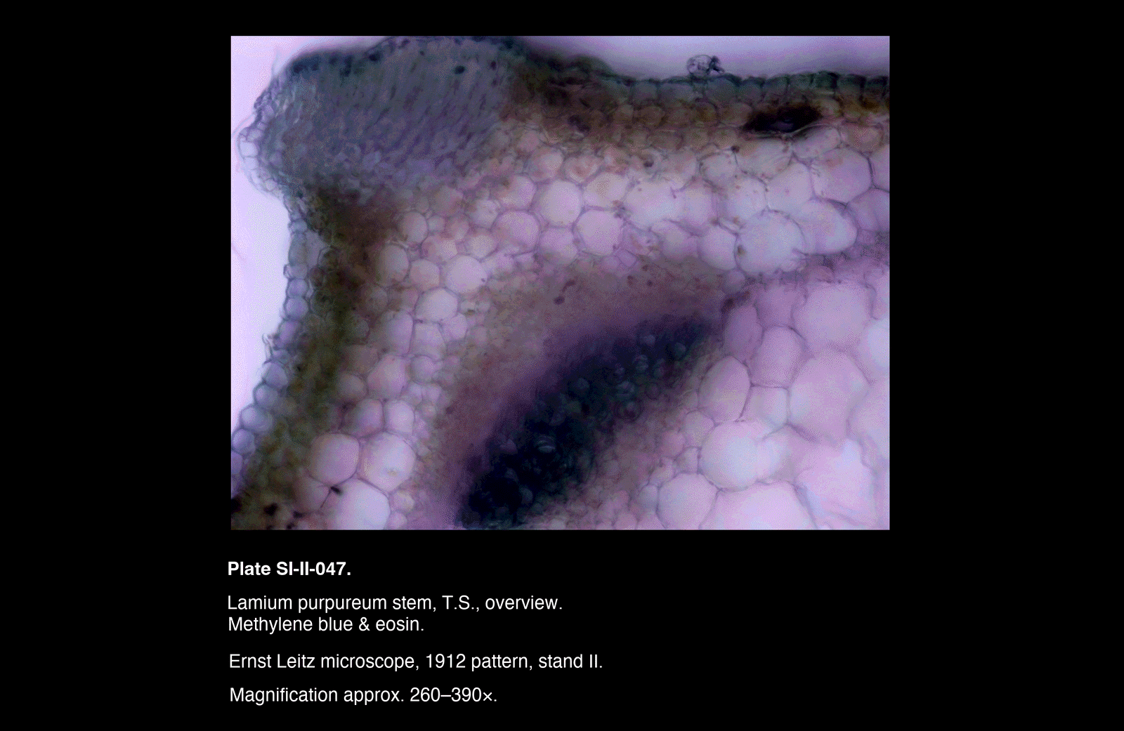
| SI-II-044 to 047 | Stained | Overview, outer, transition |
Plates
/Lamium%20purpureum%20stem,%20T.S.,%20outer%20region)
/Lamium%20purpureum%20stem,%20T.S.,%20outer%20region)
/Lamium%20purpureum%20stem,%20T.S.,%20overview)
/Lamium%20purpureum%20stem,%20T.S.,%20outer%20region)
/Lamium%20purpureum%20stem,%20T.S.,%20detail)
/Lamium%20purpureum%20stem,%20T.S.,%20overview)
/Lamium%20purpureum%20stem,%20T.S.,%20outer%20region)
/Lamium%20purpureum%20stem,%20T.S.,%20transitional%20zone)
/Lamium%20purpureum%20stem,%20T.S.,%20overview)
Plates
Observations
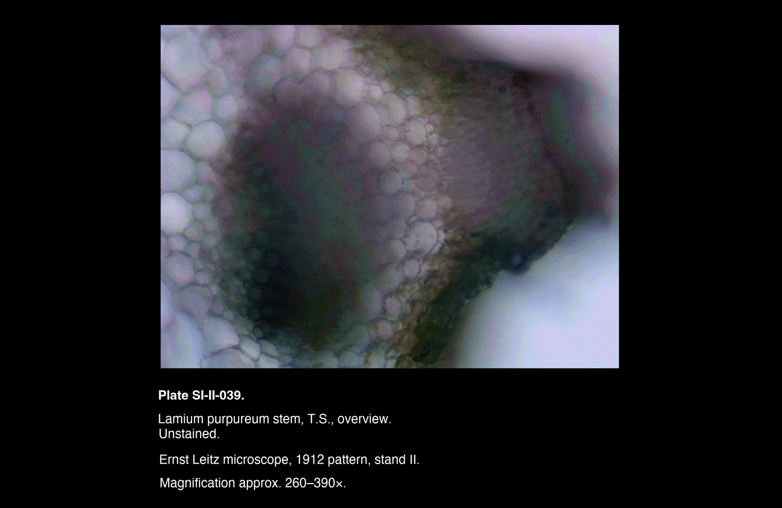
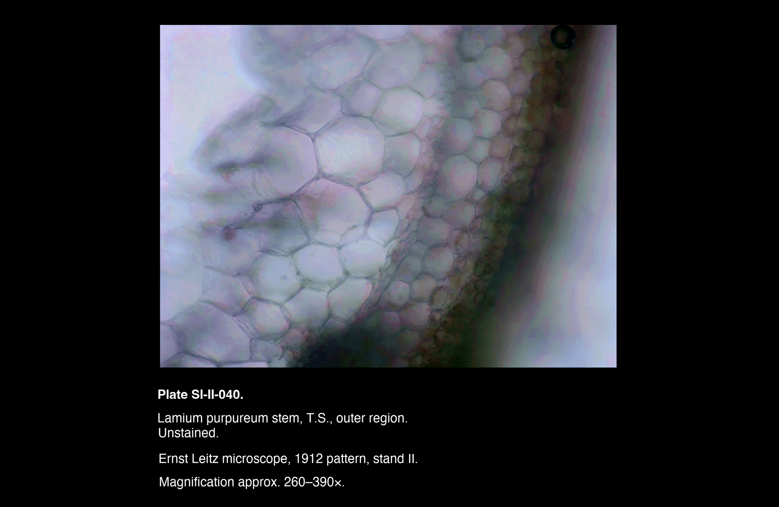
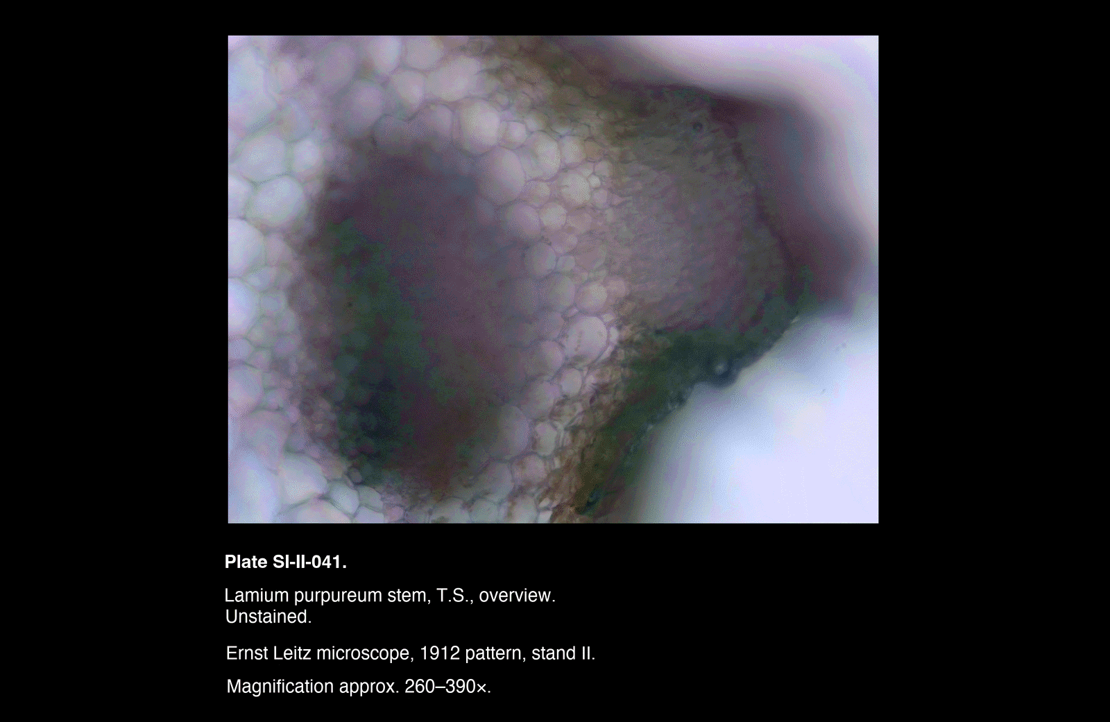
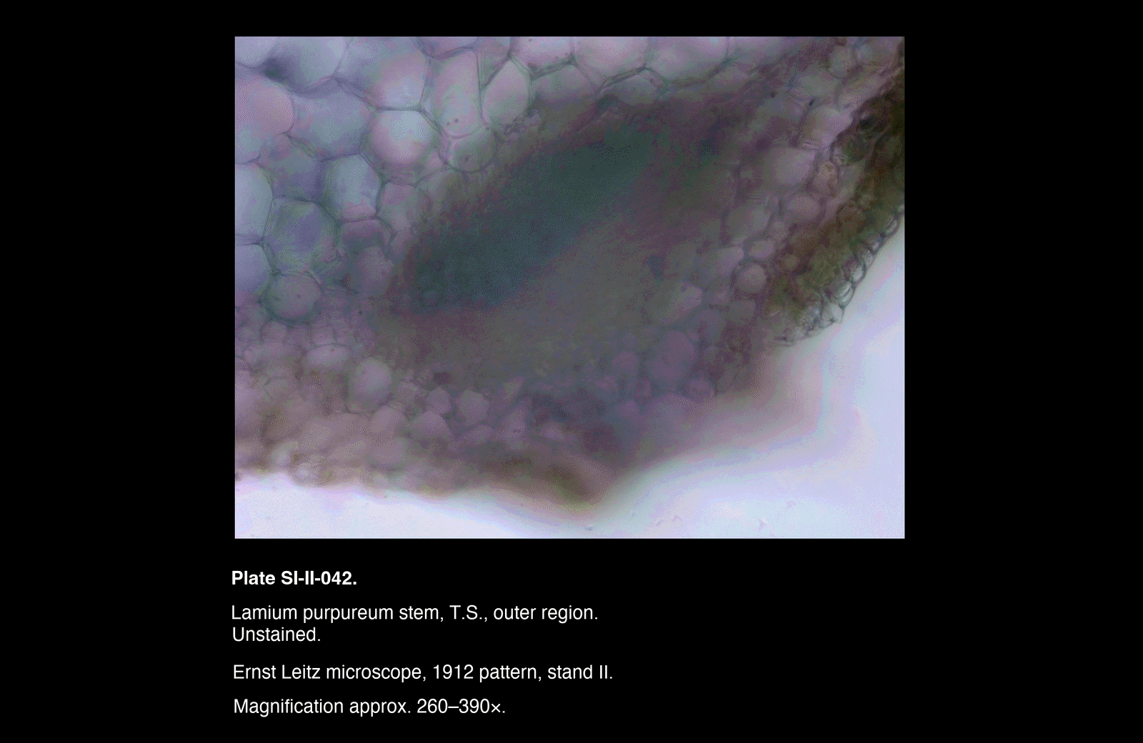
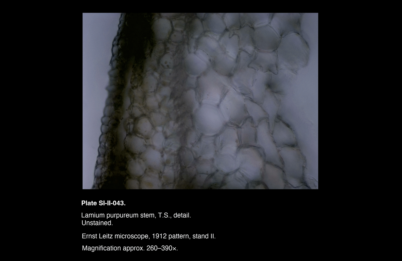
Unstained Sections (SI-II-039 → 043)
- Tissue structure visible primarily through natural contrast
- Cell walls faint but discernible
- Parenchymatous regions appear pale and relatively uniform
- Vascular regions visible but poorly differentiated from surrounding tissue
- Overall image soft, with limited contrast between tissue types
Detail views (e.g. SI-II-043) show:
- Cellular boundaries present but low in definition
- Limited internal differentiation within vascular areas
- Outer tissue boundary visible but not strongly emphasised
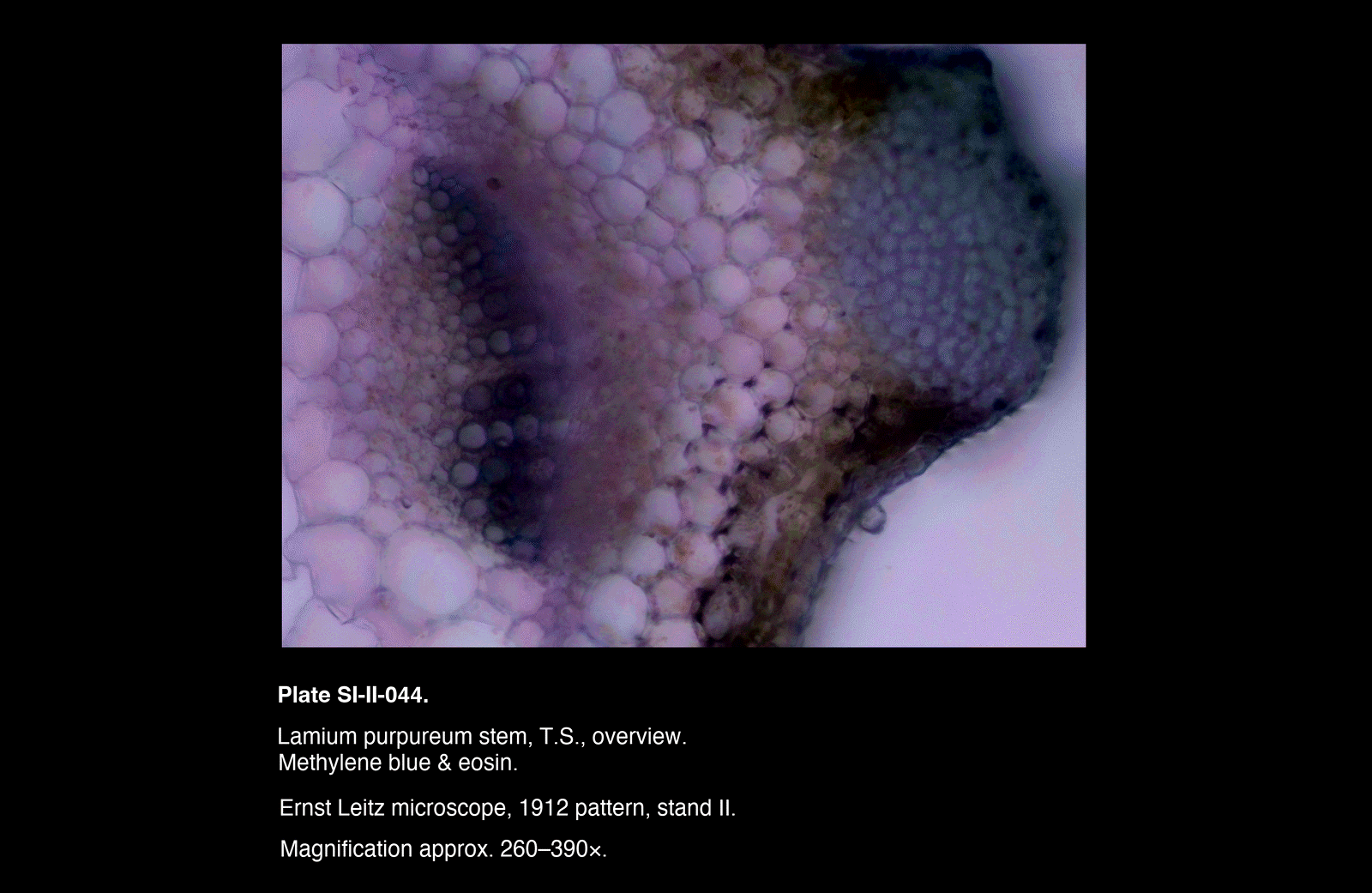
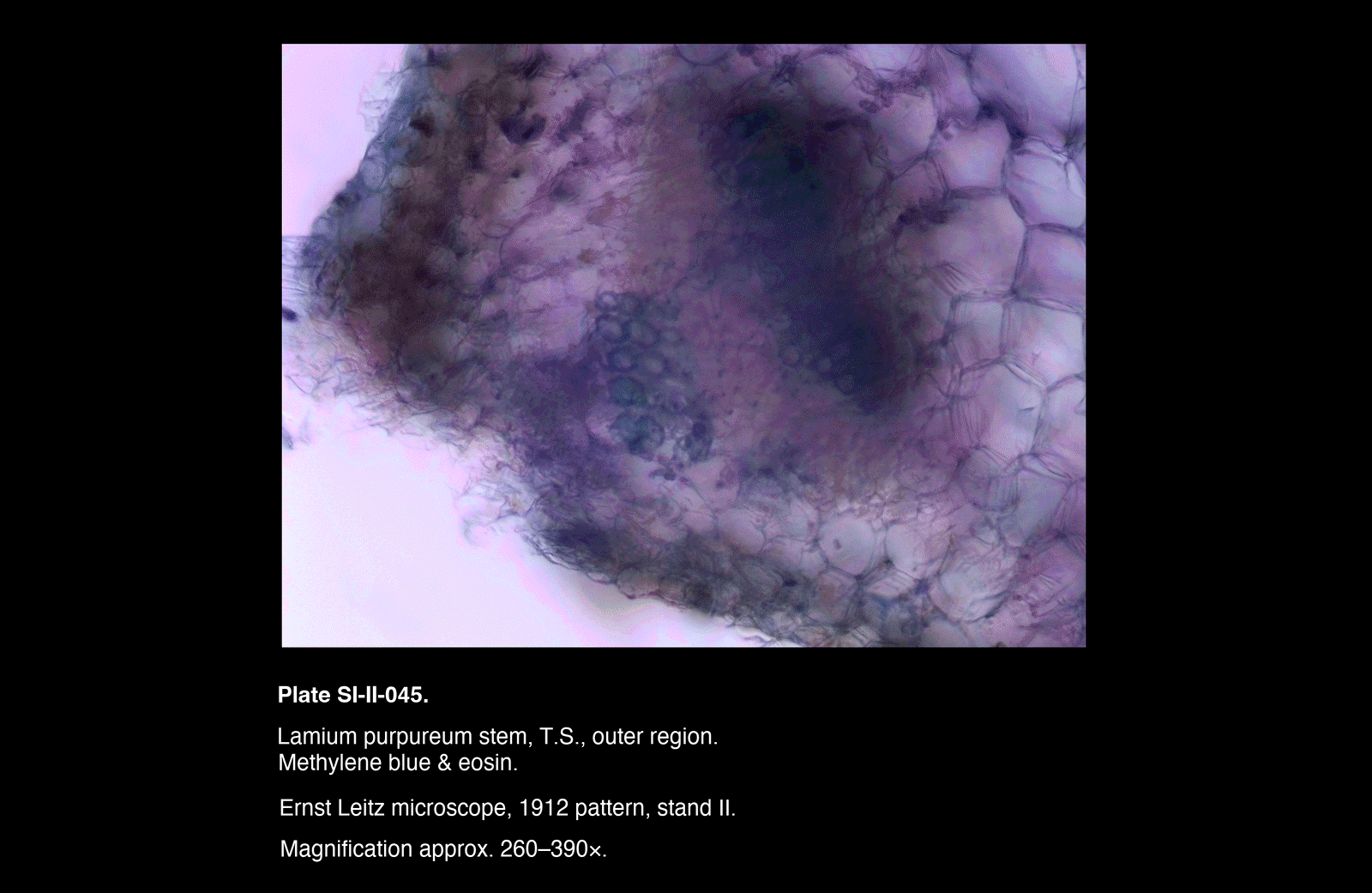
Stained Sections (SI-II-044 → 047)
- Marked increase in contrast across all regions
- Vascular bundles show strong uptake of stain, appearing distinctly darker
- Outer tissues more clearly delineated
- Parenchyma retains lighter tone, improving structural separation
In particular:
- Methylene blue appears to emphasise denser or more structured regions
- Eosin contributes a broader background tint, enhancing overall readability
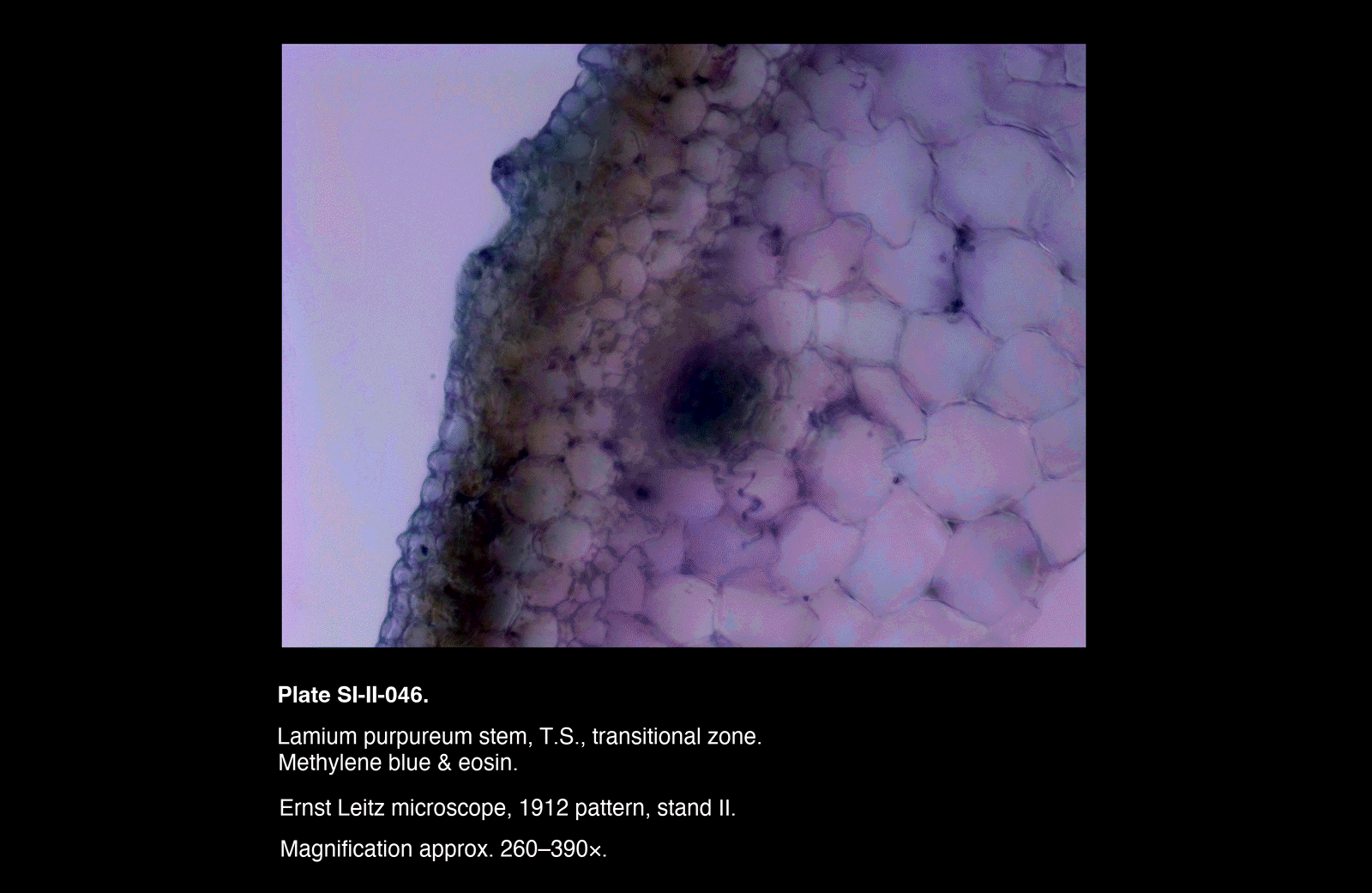
- Transitional zones (e.g. SI-II-046) become more apparent than in unstained sections
Comparative Behaviour
- Staining significantly improves visibility of:
- Vascular structure
- Tissue boundaries
- Regional organisation within the stem
-
However:
- Some loss of subtle detail in heavily stained regions
- Slight reduction in transparency compared to unstained preparations
- Occasional uneven uptake likely due to section thickness variation
Interpretation
Value of Staining
The methylene blue & eosin combination functions effectively as a general contrast-enhancing stain for plant stem material.
- Unstained sections preserve natural appearance but lack clarity
- Stained sections sacrifice some subtlety in exchange for structural legibility
The method is therefore particularly useful for:
- Initial structural interpretation
- Demonstrating tissue organisation
- Producing illustrative plates
Limitations
- Stain uptake is not highly selective
- Results are sensitive to:
- Section thickness
- Staining duration
- Rinsing consistency
- Fine intracellular detail is not substantially enhanced
Practical Outcome
This investigation establishes a reliable baseline approach for routine staining of soft plant tissues using:
- Simple reagents
- Minimal preparation steps
- Consistent visual improvement over unstained material
Remarks
- Variation between plates reflects both staining behaviour and differences in section thickness
- The inclusion of both overview and regional/detail plates proved useful in assessing the effect of staining at multiple scales
- Transitional zones (SI-II-046) are particularly informative and may merit targeted study in future work